How Cancer Spreads: The Biology Behind Metastasis
Metastasis unfolds as a chain of demanding biological steps — from escaping the original tumor to surviving the bloodstream and sometimes lying dormant for years before forming a new tumor.
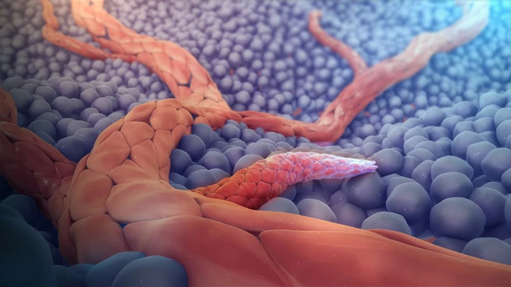
Key Takeaways
- •Metastasis is not one event but a multistep process: local invasion, entry into a vessel, survival in circulation, exit into new tissue, and growth at the new site.
- •Epithelial-mesenchymal transition (EMT) helps cancer cells lose their normal stickiness and gain the mobility needed to invade nearby tissue, partly through loss of the adhesion protein E-cadherin.
- •Tumor growth beyond a small size requires angiogenesis, the formation of new blood vessels, which also creates a route for cancer cells to enter circulation.
- •The vast majority of circulating tumor cells die before they can form a new tumor, making successful metastasis a highly inefficient process.
- •Certain cancers preferentially spread to specific organs, a pattern described over a century ago as the 'seed and soil' hypothesis and still supported by modern research.
- •Primary tumors may send signals ahead of time that prepare distant organs for incoming cancer cells, a concept called the pre-metastatic niche, though this remains an active research area.
- •Cells that reach a new organ may reverse their earlier mobility program to help them settle, suggesting EMT is a flexible, reversible state rather than a one-way switch.
- •Some disseminated cancer cells enter a dormant state and can remain inactive for years before reactivating, which helps explain late cancer recurrence.
Most people who die from cancer are not killed by the original tumor itself. They are killed by what that tumor sends elsewhere. This spread, called metastasis, is why cancer staging matters so much, and why a contained, early tumor behaves so differently from one that has already reached the liver, lungs, or bone. Metastasis is now considered one of the defining features that separates a cancer cell from an ordinary one, alongside traits like uncontrolled growth. Getting from one tumor to a second, distant one is not a single event. It is a long chain of separate steps, and a cell has to succeed at every one of them.
Why Spreading Defines Cancer's Danger
Uncontrolled growth explains why a tumor forms in the first place. It does not explain why a cancer becomes life-threatening. That distinction usually comes down to whether cells have left the original site and taken hold somewhere else. Invasion and the capacity to spread are treated as a defining requirement for cancer to progress beyond a local disease. The full sequence runs from local invasion, to entry into a blood or lymph vessel, to survival during that hostile journey, to exit into new tissue, and finally to growth there into a detectable tumor. Skip any one of these steps, and the disease stays contained.
Breaking Away From the Original Tumor
Cells inside a normal tissue are anchored, held tightly to their neighbors by adhesion proteins that keep the tissue organized. One of the most important is E-cadherin, a protein that works like molecular glue between epithelial cells, the type that lines organs such as the breast, lung, or colon. Many epithelial cancers lose this glue as they become more invasive. That loss is closely tied to a broader cellular program called epithelial-mesenchymal transition, or EMT. During EMT, a cancer cell trades its settled, structured identity for a looser, more mobile one, somewhat like a fixed shop that suddenly gains the ability to pack up and move. Cells that undergo this shift become better at pushing through the surrounding tissue and squeezing into nearby vessels. EMT is considered one of the earliest and most consequential steps in the spreading process, though how completely, and how reversibly, any given cell undergoes it likely varies.
Why New Blood Vessels Make the Escape Possible
Tumors hit a physical limit early on: cells more than a fraction of a millimeter from a blood vessel cannot get enough oxygen and nutrients to keep growing. Overcoming that limit requires angiogenesis, the growth of new blood vessels into the tumor, a process whose importance was first laid out in Judah Folkman's foundational work in the early 1970s. Angiogenesis solves the growth problem, but it creates a second one. It also builds the very route cancer cells need to leave. A tumor's new, often disorganized vessels give cells a way into the bloodstream from tissue that would otherwise contain them. Growth and spread turn out to depend on the same underlying process, rather than being two separate problems.
The Long Odds of the Journey
Once cancer cells enter the bloodstream, they become circulating tumor cells, and their situation gets precarious fast. Blood flow, immune activity, and the physical stress of being unattached in a vessel kill the overwhelming majority of them. Metastasis has been described as a strikingly inefficient process for exactly this reason: only a tiny fraction of circulating tumor cells that leave a tumor ever survive long enough to exit a vessel, let alone grow into a new tumor. A cell that does survive still has to exit into a new tissue and then divide repeatedly in an environment nothing like the one it came from. Most fail at that final stage too, a bit like a seed landing in a forest, where only a few land somewhere with the right conditions to sprout.
Not Just Anywhere: Why Some Organs Are Favored Targets
Cancers do not spread randomly. Certain cancers show a strong tendency to appear later in particular organs, far more often than blood flow alone would predict. This pattern is old news in one sense. It was first documented in 1889 by the surgeon Stephen Paget, who studied autopsy records of women with breast cancer and noticed secondary tumors clustering in specific organs. He proposed what is still called the "seed and soil" idea: a circulating cancer cell, the seed, can only grow where the local tissue, the soil, happens to support it. More than a century of later research, including work formally revisiting the idea in the early 2000s, has reinforced that basic picture while adding detail about what actually makes a tissue hospitable or not. This organ preference is one reason a cancer's likely pattern of spread can sometimes be anticipated before it happens.
Preparing the Ground Before Cancer Cells Arrive
More recent research has pushed the seed-and-soil idea further, suggesting the soil is not simply found, but may be partly prepared in advance. Signals released by a primary tumor appear able to travel to distant organs and alter tissue there, in some experiments even before any cancer cell arrives, creating what researchers call a pre-metastatic niche. One proposed mechanism involves bone-marrow-derived cells recruited to a future metastatic site, seemingly primed by the tumor's distant signaling to make that site more receptive. Another involves tiny signal-carrying packages released by tumor cells that travel to distant tissue and, in some experimental models, appear able to redirect where a cancer later spreads. Both ideas come mainly from preclinical and mechanistic studies, so they are best understood as an active, evolving area of research rather than settled clinical fact.
Settling In, and Sometimes Waiting for Years
A circulating tumor cell that survives the trip and exits into new tissue is not finished adapting. Many appear to reverse the mobility program that got them there, regaining the stickiness and structure of a settled epithelial cell. That reversal may help a stray, mobile cell behave more like the cells that normally build organs. It is further evidence that the mobile state cancer cells adopt early in spreading is not a one-way switch; cells seem able to move into it and back out of it depending on what a given stage of the journey requires, though the process observed in patients is likely more varied than any simplified model captures.
Not every cell that reaches a new organ grows right away. Some slip into a dormant state, alive but not dividing, sometimes for years. This tumor dormancy helps explain one of metastasis's more unsettling features: cancer that returns long after treatment appeared to succeed, occasionally a decade or more later. What keeps a dormant cell inactive, and what eventually triggers it to grow, is still only partly understood. Researchers are actively mapping the signals involved, but timing and triggers are not yet predictable for any individual patient.
An Unfinished Picture With Real Stakes
Taken together, metastasis looks less like a single event and more like an obstacle course. A cancer cell has to detach, survive, land somewhere accommodating, and sometimes wait in silence before it ever becomes a second tumor a scan can detect. Every stage described here, invasion, circulation, organ targeting, niche preparation, and dormancy, is a point where the process usually fails, which is exactly why metastasis is rare relative to how many cells a tumor sheds. It is also why these stages are studied separately: whatever lets a cell escape a tumor is not necessarily what lets it survive elsewhere, or what eventually wakes it up. Much of this remains a live area of research, particularly the earliest distant-organ-priming signals and the biology of dormancy, and firm conclusions about timing or triggers in real patients are still out of reach.
Common Questions
References
- Ann F. Chambers, Alan C. Groom, Ian C. MacDonald (2002). Dissemination and growth of cancer cells in metastatic sites
- Scott Valastyan, Robert A. Weinberg (2011). Tumor Metastasis: Molecular Insights and Evolving Paradigms
- Jean Paul Thiery, Hervé Acloque, Ruby Y.J. Huang, M. Angela Nieto (2009). Epithelial-Mesenchymal Transitions in Development and Disease
- Raghu Kalluri, Robert A. Weinberg (2009). The basics of epithelial-mesenchymal transition
- Jing Yang, Robert A. Weinberg (2008). Epithelial-Mesenchymal Transition: At the Crossroads of Development and Tumor Metastasis
- Joan Massagué, Anna C. Obenauf (2016). Metastatic colonization by circulating tumour cells
- Stephen Paget (1889). THE DISTRIBUTION OF SECONDARY GROWTHS IN CANCER OF THE BREAST.
- Isaiah J. Fidler (2003). The pathogenesis of cancer metastasis: the 'seed and soil' hypothesis revisited
- Héctor Peinado, Haiying Zhang, Irina R. Matei, Bruno Costa-Silva, Ayuko Hoshino, Goncalo Rodrigues, Bethan Psaila, Rosandra N. Kaplan, Jacqueline F. Bromberg, Yibin Kang, Mina J. Bissell, Thomas R. Cox, Amato J. Giaccia, Janine T. Erler, Sachie Hiratsuka, Cyrus M. Ghajar, David Lyden (2017). Pre-metastatic niches: organ-specific homes for metastases
- (2005). VEGFR1-positive haematopoietic bone marrow progenitors initiate the pre-metastatic niche.
- Héctor Peinado, Maša Alečković, Simon Lavotshkin, Irina Matei, Bruno Costa-Silva, Gema Moreno-Bueno, Marta Hergueta-Redondo, Caitlin Williams, Guillermo García-Santos, Cyrus M Ghajar, Ayuko Nitadori-Hoshino, Caitlin Hoffman, Karen Badal, Benjamin A Garcia, Margaret K Callahan, Jianda Yuan, Vilma R Martins, Johan Skog, Rosandra N Kaplan, Mary S Brady, Jedd D Wolchok, Paul B Chapman, Yibin Kang, Jacqueline Bromberg, David Lyden (2012). Melanoma exosomes educate bone marrow progenitor cells toward a pro-metastatic phenotype through MET
- (2014). Mechanisms of disseminated cancer cell dormancy: an awakening field.
- Louis M. Sherwood, Edith E. Parris, Judah Folkman (1971). Tumor Angiogenesis: Therapeutic Implications
- Douglas Hanahan, Robert A. Weinberg (2011). Hallmarks of Cancer: The Next Generation
Team Review: This article has been reviewed by Canceria Research Team