What Makes Multiple Myeloma Different From Other Blood Cancers
Why a cancer of mature plasma cells follows its own path in biology, diagnosis, and treatment compared with leukemia and lymphoma
Key Takeaways
- •Multiple myeloma arises from mature plasma cells, unlike leukemia, which starts in immature blood cells, or lymphoma, which starts in lymphocytes that form tumors.
- •The World Health Organization classifies myeloma alongside lymphoma as a lymphoid neoplasm, reflecting shared cell lineage despite very different clinical behavior.
- •Nearly all multiple myeloma cases are preceded by MGUS, a common and usually harmless condition that is monitored over time rather than treated immediately.
- •Myeloma is marked by recurrent chromosomal translocations and complex karyotypes that are not typical features of most leukemias.
- •Myeloma uniquely disrupts the normal balance between bone-building and bone-breaking cells, producing osteolytic bone lesions rarely seen in other blood cancers.
- •Diagnosis relies on the CRAB criteria and detection of an abnormal M protein, which differ substantially from how leukemia and lymphoma are diagnosed.
- •Myeloma treatment centers on proteasome inhibitors, immunomodulatory drugs, and newer BCMA-targeted therapies, distinct from standard leukemia and lymphoma regimens.
- •Multiple myeloma incidence is roughly twice as high in Black populations as in white populations, a disparity larger than seen in most other blood cancers.
"Blood cancer" is really an umbrella term for diseases with different origins. Multiple myeloma is one of those diseases, but it differs from leukemia and lymphoma in a way that reaches deep into basic biology. Understanding that difference explains why myeloma causes bone pain and fractures, why it is diagnosed differently, and why it is treated with its own set of drugs.
A Different Starting Cell
Each major blood cancer traces back to a different kind of blood cell gone wrong. Leukemia typically starts in young, developing blood cells still inside the bone marrow, cells that have not finished maturing and often spill into the bloodstream in large numbers. Lymphoma begins in lymphocytes, immune cells that usually travel through lymph nodes and other lymphoid tissue, where they can form solid tumor masses.
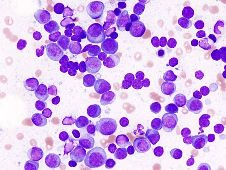
Multiple myeloma starts somewhere else entirely: in plasma cells. A plasma cell is a mature white blood cell whose normal job is producing antibodies, the proteins the immune system uses to recognize and fight infection. In myeloma, a single plasma cell becomes malignant and multiplies, flooding the bone marrow with copies of itself and churning out a single, abnormal antibody fragment called an M protein, detectable through blood or urine tests. Unlike leukemia cells, myeloma cells rarely spill into general circulation in large numbers. Unlike lymphoma, they rarely form the kind of solid tumor mass found in a lymph node. They stay put in the bone marrow, which turns out to matter enormously for how the disease behaves.
Where Myeloma Fits in the Official Classification
Blood cancers are formally organized by the World Health Organization into broad categories based on which cell lineage and developmental stage a cancer arises from. One major branch covers myeloid neoplasms, including most leukemias, which arise from blood-forming cells that would normally become red cells, platelets, or certain white cells. A separate branch covers lymphoid neoplasms, which includes lymphoma and, perhaps surprisingly, multiple myeloma itself, grouped there as a plasma cell neoplasm.
That placement reflects lineage rather than appearance. Plasma cells are the mature end stage of B lymphocyte development, the same broad immune cell family that gives rise to many lymphomas. Myeloma and lymphoma are molecular cousins in that sense, even though the two diseases look and behave quite differently in a patient.
A Long, Quiet Precursor Phase
Few cancers announce their precursor state as clearly as multiple myeloma does. Almost every case is preceded, often for years or decades, by a condition called monoclonal gammopathy of undetermined significance, or MGUS. In MGUS, abnormal plasma cells are already present and already producing a detectable M protein, but the person has no symptoms and no organ damage. A landmark long-term study led by Robert Kyle and colleagues tracked people with MGUS for decades and found that it converts to myeloma or a related cancer at a low, fairly steady rate each year, rather than all at once.
That slow, predictable background rate is unusual among blood cancer precursors. It is also why MGUS is something doctors monitor rather than treat. Having MGUS does not mean a person has cancer; it means they carry a small, ongoing chance of developing it later, which is why routine follow-up blood tests, not immediate treatment, are the standard response.
A Genetic Signature of Its Own
At the molecular level, myeloma cells tend to carry a distinctive pattern of damage. Many cases involve a broken piece of chromosome swapping places with the region of DNA that normally instructs antibody-producing cells how to build antibodies. These rearrangements, along with a generally more chaotic chromosome count than is typical in most leukemias, appear early and help shape how the disease develops, according to molecular studies of myeloma genetics. This genomic complexity is one reason myeloma is now understood less as a single disease and more as a collection of molecularly distinct subtypes that happen to share a cell of origin and a clinical picture.
Why the Skeleton Becomes a Target
Few features separate myeloma from other blood cancers as sharply as its effect on bone. Healthy bone is in constant quiet renewal, with one type of cell dissolving old bone and another rebuilding it, a balance kept in careful check. Myeloma cells disrupt that balance directly: they push the bone-dissolving side into overdrive while suppressing the bone-building side, producing what are called osteolytic lesions, areas where bone is destroyed faster than it can be replaced.
The result is a disease that commonly causes bone pain, thinning, and fractures, something rarely seen as a defining feature of leukemia or lymphoma. This is reflected directly in how myeloma is diagnosed: doctors look for a set of features known by the shorthand CRAB, standing for elevated calcium, reduced kidney function, anemia, and bone lesions, alongside the presence of the M protein and other markers. Leukemia diagnoses instead hinge on counting abnormal cells in blood or marrow, and lymphoma diagnoses typically rely on biopsy of an enlarged lymph node or other tissue mass. Three different diseases, three genuinely different diagnostic playbooks. Recognizing these patterns is not the same as diagnosing them; that still requires laboratory testing and clinical evaluation by a qualified physician.
A Disease That Depends on Its Neighborhood
Myeloma cells are unusually dependent on the bone marrow microenvironment, the supportive mesh of cells and tissue that normally surrounds developing blood cells. Think of it less like a tumor growing on its own and more like a tenant that cannot survive outside a very specific apartment; myeloma cells lean on signals from their marrow neighbors to stay alive, grow, and resist damage. That dependence also helps explain the bone destruction described above, since some of the same marrow signals that myeloma cells exploit for survival are the ones that tip the bone-remodeling balance out of place.
Different Biology, Different Drug Classes
Because myeloma's vulnerabilities are so distinct, its treatment has evolved around drug classes rarely central to leukemia or lymphoma care. Proteasome inhibitors block a structure inside the cell responsible for breaking down and recycling proteins, a process myeloma cells rely on heavily given how much antibody protein they churn out. Immunomodulatory drugs and antibodies that target a specific marker on plasma cells round out a now-standard combination approach.
More recently, therapies aimed at a protein called BCMA, found mainly on plasma cells including myeloma cells, have entered clinical use for people whose disease has returned after other treatments. One such approach, a form of engineered immune cell therapy, showed meaningful responses in a clinical trial of patients with relapsed and refractory disease, though it is reserved for specific situations rather than offered as a universal or guaranteed solution. These drug classes have little overlap with standard leukemia or lymphoma regimens, a direct consequence of starting from such a different cell of origin.
Who Gets Myeloma, and How That Differs Too
Multiple myeloma also shows a distinct population pattern. Research led by Adam Waxman and colleagues found that myeloma occurs roughly twice as often in Black populations as in white populations in the United States, a disparity notably larger than seen in most other blood cancers. The reasons behind that gap are not fully settled, and the finding describes a pattern across large populations, not a prediction for any individual person.
A Disease With Its Own Rules
Multiple myeloma shares the label "blood cancer" with leukemia and lymphoma, but little else lines up neatly. It starts in a different, more mature cell. It is classified alongside lymphoma on a technicality of lineage, while behaving nothing like it clinically. It spends years announcing itself quietly through MGUS before anything resembling active cancer appears. Its genetics, its effect on bone, its treatment, and even who it affects most, all trace back to one central fact: myeloma is fundamentally a disease of the plasma cell, and almost everything distinctive about it follows from that single starting point.
Common Questions
References
- Robert A. Kyle, Terry M. Therneau, S. Vincent Rajkumar, Janice R. Offord, Dirk R. Larson, Matthew F. Plevak, L. Joseph Melton (2002). A Long-Term Study of Prognosis in Monoclonal Gammopathy of Undetermined Significance
- P. Leif Bergsagel, W. Michael Kuehl (2005). Molecular Pathogenesis and a Consequent Classification of Multiple Myeloma
- Gareth J. Morgan, Brian A. Walker, Faith E. Davies (2012). The genetic architecture of multiple myeloma
- G. David Roodman (2009). Pathogenesis of myeloma bone disease
- Nikhil C. Munshi, Larry D. Anderson, Nina Shah, Deepu Madduri, Jesús Berdeja, Sagar Lonial, Noopur Raje, Yi Lin, David Siegel, Albert Oriol, Philippe Moreau, Ibrahim Yakoub-Agha, Michel Delforge, Michele Cavo, Hermann Einsele, Hartmut Goldschmidt, Katja Weisel, Alessandro Rambaldi, Donna Reece, Fabio Petrocca, Monica Massaro, Jamie N. Connarn, Shari Kaiser, Payal Patel, Liping Huang, Timothy B. Campbell, Kristen Hege, Jesús San-Miguel (2021). Idecabtagene Vicleucel in Relapsed and Refractory Multiple Myeloma
- Adam J. Waxman, Pamela J. Mink, Susan S. Devesa, William F. Anderson, Brendan M. Weiss, Sigurdur Y. Kristinsson, Katherine A. McGlynn, Ola Landgren (2010). Racial disparities in incidence and outcome in multiple myeloma: a population-based study
- Steven H. Swerdlow, Elias Campo, Stefano A. Pileri, Nancy Lee Harris, Harald Stein, Reiner Siebert, Ranjana Advani, Michele Ghielmini, Gilles A. Salles, Andrew D. Zelenetz, Elaine S. Jaffe (2016). The 2016 revision of the World Health Organization classification of lymphoid neoplasms
Team Review: This article has been reviewed by Canceria Research Team