Why Mammograms and Pap Smears Work So Differently
How the underlying biology of cervical and breast cancer shaped two very different approaches to catching disease early
Key Takeaways
- •A screening test looks for disease in people with no symptoms, while a diagnostic test confirms or rules out disease once something has already raised suspicion.
- •Persistent infection with high-risk HPV is a necessary cause of nearly all cervical cancer, giving it an unusually identifiable trigger compared with most other cancers.
- •Cervical cancer passes through a long, well-characterized precancerous stage called cervical intraepithelial neoplasia, which can be detected years before invasive cancer develops.
- •Because the cervix naturally sheds surface cells, Pap smear cytology can directly examine those cells for early abnormal changes, an approach first described in the 1940s.
- •Population-based cervical cytology screening programs have been linked to substantial reductions in cervical cancer incidence and mortality.
- •Breast cancer lacks an accessible shedding surface and a single well-defined precancerous stage, which is why mammography relies on X-ray imaging of structural changes instead of cell sampling.
- •Breast density can obscure tumors on a mammogram, lowering the test's sensitivity in denser breast tissue.
- •All screening involves tradeoffs, including false positives, overdiagnosis, and statistical illusions like lead-time and length-time bias that can make early detection look more beneficial than it truly is.
A Pap smear and a mammogram are both described as cancer screening tests, but they don't look for the same thing, and they don't work the same way. One collects cells from the surface of the cervix and studies them under a microscope. The other takes an X-ray picture of the whole breast, looking for a mass or a telltale cluster of calcium deposits. That difference isn't a matter of preference or available technology. It comes directly from how each cancer forms.
What Makes a Test a "Screening" Test
A screening test is given to people who feel completely fine, with no symptoms at all. Its job is to catch disease before it would otherwise announce itself. A diagnostic test is different: it's used once something has already raised suspicion, whether that's a symptom, a lump, or an abnormal screening result. Screening has to work at a population scale, on mostly healthy people, which means it has to be simple, repeatable, and reasonably cheap. Those practical constraints matter as much as the biology does, but biology decides what the test is actually looking for.
A Virus With a Visible Fingerprint
Cervical cancer has an unusual feature that makes it especially well suited to screening. Nearly all cases are caused by persistent infection with certain high-risk types of human papillomavirus, or HPV, a common virus that most sexually active people encounter at some point. In most people the immune system clears the infection. In a smaller number, the virus lingers for years, and that persistence is what drives the cervix's surface cells to accumulate the kind of damage that eventually becomes cancer. This causal relationship is about as close to a known, singular trigger as cancer research gets, which is unusual: most cancers don't have one identifiable cause you can point to.
Because HPV acts slowly and visibly, the cervix passes through a recognizable in-between stage before cancer develops. Pathologists call this cervical intraepithelial neoplasia, often shortened to CIN: cells that look abnormal under a microscope but aren't yet cancerous. This stage can last for years, sometimes over a decade, giving doctors a long window to intervene. Few other cancers offer that kind of advance warning.
Catching Abnormal Cells Before They Become Cancer
This is exactly what makes cytology, the study of individual cells, such a good match for cervical screening. The cervix sheds cells naturally, and a sample can be collected with a simple swab during a routine exam. A pathologist then looks at those cells directly, checking for the shape and structure changes that mark early abnormality. This approach dates back to the 1940s, when George Papanicolaou and his colleague Herbert Traut first showed that abnormal cervical cells could be identified in a simple vaginal smear, work that gave the Pap smear its name. The underlying idea hasn't changed much since: find the abnormal cells before they turn into something invasive.
The real-world impact of that idea has been substantial. Where cytology-based cervical screening programs have been rolled out at a population level, cervical cancer incidence and deaths have dropped sharply compared with what models predict would otherwise have occurred. Analyses of screening in the United Kingdom, for example, estimated that organized Pap testing prevented a cervical cancer epidemic that would otherwise have developed as HPV infection became more common. That's a rare case of a simple, decades-old test producing a clear, measurable public health effect.
Why Breast Tissue Needs a Different Kind of Test
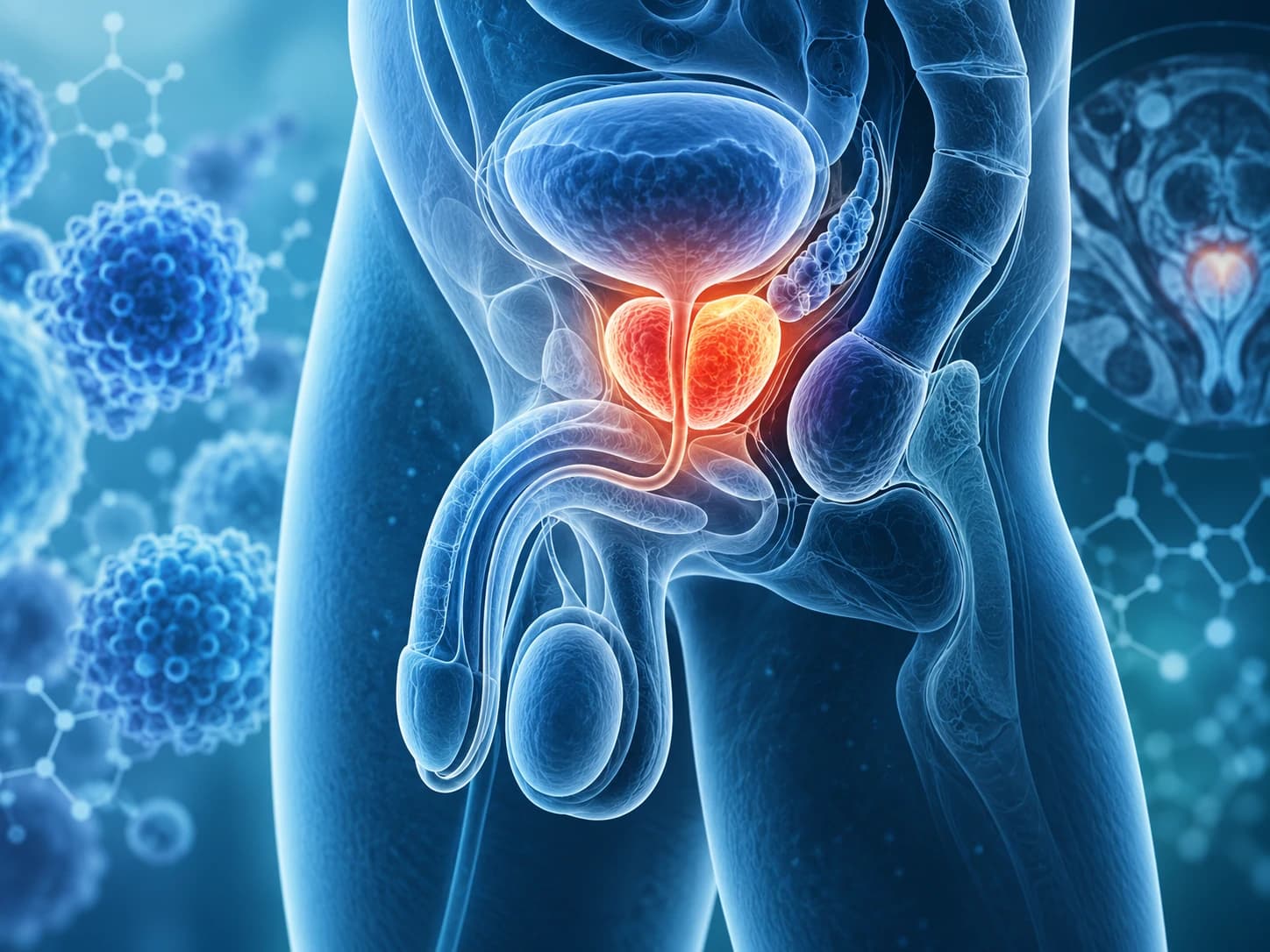
Breast cancer doesn't offer anything quite like that visible, cell-shedding surface. The cervix is a passage lined with a layer of cells that naturally flake off and can be collected with a swab. The breast is solid tissue, buried under skin and fat, with no equivalent surface to sample directly. There's also no single, well-defined precancerous stage in the breast that plays the same role CIN plays in the cervix, detectable years in advance from a scraping of surface cells. So screening has to work from the outside in, which is exactly what mammography does.
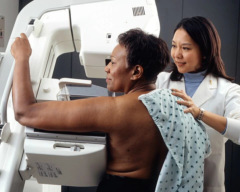
Mammography is an X-ray technique that looks for structural clues rather than cellular ones: a dense mass that doesn't belong, or a pattern of tiny calcium deposits that can mark early changes. It's less like reading a cell under a microscope and more like looking at a photograph for something that doesn't fit the background. That analogy also explains one of mammography's real limitations. Breast density, the amount of fibrous and glandular tissue relative to fat, can make a tumor and the surrounding tissue look similarly pale on the image, the same way a detail is harder to spot the more cluttered the photograph. In denser breasts, that overlap can lower a mammogram's sensitivity, its ability to correctly flag disease that's actually there.
The Hidden Costs of Looking Early
No screening test is free of tradeoffs, and the tradeoffs follow directly from what sensitivity and specificity mean in practice. A highly sensitive test catches more true cases, but it also tends to flag more things that turn out not to be cancer, lowering specificity, the test's ability to correctly clear people who are disease-free. Every screening program has to balance those two properties, and neither test gets it perfectly.
Screening also introduces subtler statistical illusions that are easy to misread as treatment success. If a cancer is found earlier, a person may appear to survive longer after diagnosis, even if the disease was always going to take the same course either way; this is called lead-time bias, and it can make early detection look more beneficial than it actually is. A related effect, sometimes called length-time bias, means that screening tends to catch slower-growing cancers more often simply because they stick around longer to be found, while fast-growing ones can appear between screenings. Together these effects can make screening look like it's finding more disease and saving more lives than it strictly is, a distortion first laid out clearly in the early 1990s in work examining how advances in imaging were overestimating both disease prevalence and the benefits of treating what imaging found. Some of what's caught through screening, particularly slow-growing abnormalities, may never have caused harm in a person's lifetime at all, a phenomenon generally referred to as overdiagnosis. None of this means screening doesn't work. It means its benefits have to be weighed carefully against its costs, rather than assumed automatically.
A Shift Already Underway in Cervical Screening
Knowing that HPV itself drives nearly all cervical cancer has reshaped how cervical screening is approached in some settings, adding or substituting direct tests for high-risk HPV types alongside, or instead of, traditional cytology. The logic is straightforward: if the virus is the necessary trigger, testing for its presence can flag risk even before any cellular abnormality appears. This doesn't replace clinical screening guidelines, which vary by country and by individual risk and are set by health authorities rather than by any single study. It does illustrate something broader, though: as understanding of a cancer's cause deepens, screening strategies built around that cancer tend to evolve with it.
Why One Test Can't Cover Every Cancer
The differences between a Pap smear and a mammogram aren't a story of one test being more advanced or more sophisticated than the other. They reflect two cancers with genuinely different biology: one with an identifiable viral cause and a long, visible precancerous runway, the other without a comparable single trigger or an accessible surface to sample from. A universal screening test would require a universal kind of cancer, and cancer simply isn't that uniform. Each test is built around the specific way its target disease reveals itself, which is also why the right screening approach, and how often to use it, is a decision made with a healthcare provider rather than a one-size-fits-all rule.
Common Questions
References
- Jan M. M. Walboomers, Marcel V. Jacobs, M. Michele Manos, F. Xavier Bosch, J. Alain Kummer, Keerti V. Shah, Peter J. F. Snijders, Julian Peto, Chris J. L. M. Meijer, Nubia Mu�oz (1999). Human papillomavirus is a necessary cause of invasive cervical cancer worldwide
- (2002). Papillomaviruses and cancer: from basic studies to clinical application.
- George N. Papanicolaou, Herbert F. Traut (1941). The Diagnostic Value of Vaginal Smears in Carcinoma of the Uterus
- Julian Peto, Clare Gilham, Olivia Fletcher, Fiona E Matthews (2004). The cervical cancer epidemic that screening has prevented in the UK
- William C. Black, H. Gilbert Welch (1993). Advances in Diagnostic Imaging and Overestimations of Disease Prevalence and the Benefits of Therapy
Team Review: This article has been reviewed by Canceria Research Team