How Melanoma Develops: From a Single Skin Cell to an Aggressive Cancer
A step-by-step look at the sun damage, gene mutations, and immune evasion that turn a pigment cell into invasive melanoma.
Key Takeaways
- •Melanocytes are pigment-producing skin cells that normally divide rarely; melanoma begins when that restraint breaks down.
- •UV radiation damages DNA through lesions like pyrimidine dimers, producing a distinctive high mutation burden in sun-exposed melanocytes.
- •About half of cutaneous melanomas carry a BRAF mutation, commonly V600E, which locks the MAPK growth pathway in the 'on' position.
- •Loss of the CDKN2A tumor suppressor gene, whether inherited or acquired, removes a key brake on cell division and contributes to melanoma.
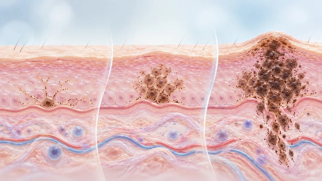
- •Melanoma can progress through a genetic staircase from benign mole to dysplastic nevus to melanoma in situ to invasive melanoma, though not every case follows this exact route.
- •As melanoma advances, tumor cells can display PD-L1 to suppress nearby immune T cells, helping the tumor evade immune detection.
- •Fair skin, high mole count, family history, and inherited genetic syndromes raise melanoma risk independent of UV exposure alone.
- •Discovering these molecular drivers led directly to targeted therapies against BRAF and immunotherapies that block checkpoint signals like PD-L1.
Skin color comes from a single kind of cell called a melanocyte, which produces the pigment that protects deeper skin layers from the sun. Normally these cells divide rarely and quietly do their job for decades. Melanoma begins when something disrupts that restraint, letting a melanocyte and its descendants multiply out of control. Tracing how that disruption happens, from the first DNA damage to a tumor that can spread, explains why melanoma looks so different from one patient to the next, and why its treatment has changed so dramatically in recent decades.
How Ultraviolet Light Damages Skin Cell DNA
UV radiation is the invisible part of sunlight, also produced by tanning beds, that carries enough energy to distort the structure of DNA. One of its signature effects is a lesion called a pyrimidine dimer, where two neighboring DNA building blocks fuse together in a way the cell's copying machinery can misread. Skin cells repair most of this damage efficiently. Still, repair is never perfect, so every unrepaired dimer carries a small risk of becoming a permanent mutation.
Over years of cumulative sun exposure, melanocytes in frequently exposed skin accumulate thousands of these small mutations. This produces a distinctive mutational pattern that researchers can recognize across melanoma genomes, a kind of molecular fingerprint of past UV damage. Most of these mutations are harmless bystanders. The danger appears when one lands inside a gene that controls how often a cell divides.
The Genetic Switch That Pushes Cells to Grow
One mutation shows up far more often than chance alone would explain: a change in the BRAF gene, most commonly a specific variant called BRAF V600E. Roughly half of all cutaneous melanomas carry a BRAF mutation like this. BRAF is one link in a relay of proteins, known as the MAPK pathway, that tells a cell when to divide.
A useful comparison is a stuck gas pedal. The mutated BRAF protein keeps sending the "grow" signal constantly, whether or not the cell actually needs to divide. Large-scale genomic studies of melanoma tumors have confirmed BRAF mutations as one of the most frequent drivers of the disease, a discovery that reshaped how researchers think about what sets melanoma in motion in the first place.
When the Brakes Fail
A stuck gas pedal becomes far more dangerous if the brakes are gone too. The CDKN2A gene normally produces a protein that holds the cell division cycle in check, stopping cells with damaged DNA from multiplying further. Losing CDKN2A function removes that restraint entirely.
This loss can be inherited, running in families with a strong history of melanoma, or it can be acquired later through new mutations within the tumor itself. Genetic studies of melanoma-prone families identified CDKN2A loss as a recurring feature, and later research showed the same gene is frequently disabled in melanomas with no family history at all. A cell carrying both a stuck BRAF switch and a disabled CDKN2A brake is in a very different situation than one with only a single hit.
From Ordinary Mole to Invasive Cancer
Most moles, or nevi, never become anything more than a mole. They form when a cluster of melanocytes picks up a single growth-related mutation, often in BRAF, and then simply stops. Some additional, still not fully understood, braking mechanism keeps most moles stable for a lifetime.
Melanoma generally requires further genetic hits layered on top of that first mutation. Detailed sequencing of tissue spanning normal skin, atypical moles known as dysplastic nevi, melanoma confined to the skin's surface layer (melanoma in situ), and invasive melanoma, work associated with Hunter Shain and Boris Bastian's research group, revealed that these stages often represent a genetic staircase. Each step up tends to carry additional mutations layered onto the ones before it.
Not every melanoma passes through a visible mole stage first, and not every dysplastic nevus goes on to progress. This staged picture describes a well-documented route, not the only possible path to melanoma.
Slipping Past the Immune System
Genetics is only part of the story, because the immune system is constantly screening for abnormal cells. As melanoma advances, some tumor cells begin displaying a protein called PD-L1 on their surface. PD-L1 works like a password: it tells nearby immune cells, specifically the T cells that would otherwise destroy abnormal cells, to stand down.
This signal is a normal safety mechanism the body uses to avoid attacking its own healthy tissue, and melanoma cells can hijack it for protection. A clinical study testing an antibody designed to block this signal, led by Suzanne Topalian and colleagues, showed that interrupting PD-L1 signaling could trigger immune responses against several cancer types, melanoma included. That finding helped establish immune checkpoint biology as a central part of how melanoma evades detection as it progresses.
Why Some People Are More Susceptible Than Others
Sun exposure is only one part of melanoma risk. Fair skin that burns easily, a high number of moles, and a family history of melanoma each raise individual risk, independent of how much UV exposure a person has had. Inherited genetic predisposition syndromes, often involving the same CDKN2A gene described earlier, can run in families and substantially raise lifetime risk.
This is why two people with similar sun exposure histories can face very different odds. Starting genetic makeup, and how many moles a person's skin has already produced over a lifetime, can matter just as much as cumulative UV dose.
How This Biology Changed Melanoma Treatment
For a long time, melanoma was treated much the same way regardless of what was happening inside individual tumor cells. Identifying BRAF mutations and PD-L1-driven immune evasion changed that. Treatments now exist that are built directly around these two mechanisms: therapies that target the overactive BRAF signal, and antibodies that block the PD-L1 signal so the immune system can recognize the tumor again.
Neither approach works for every patient, and tumors can sometimes adapt around a single blocked pathway, which is part of why research into melanoma's underlying biology remains active. What has changed permanently is the starting assumption: melanoma is no longer approached as one uniform disease, but as a condition whose behavior depends on which genetic switches and immune evasion tricks a particular tumor has acquired along the way.
Common Questions
References
- Helen Davies, Graham R. Bignell, Charles Cox, Philip Stephens, Sarah Edkins, Sheila Clegg, Jon Teague, Hayley Woffendin, Mathew J. Garnett, William Bottomley, Neil Davis, Ed Dicks, Rebecca Ewing, Yvonne Floyd, Kristian Gray, Sarah Hall, Rachel Hawes, Jaime Hughes, Vivian Kosmidou, Andrew Menzies, Catherine Mould, Adrian Parker, Claire Stevens, Stephen Watt, Steven Hooper, Rebecca Wilson, Hiran Jayatilake, Barry A. Gusterson, Colin Cooper, Janet Shipley, Darren Hargrave, Katherine Pritchard-Jones, Norman Maitland, Georgia Chenevix-Trench, Gregory J. Riggins, Darell D. Bigner, Giuseppe Palmieri, Antonio Cossu, Adrienne Flanagan, Andrew Nicholson, Judy W. C. Ho, Suet Y. Leung, Siu T. Yuen, Barbara L. Weber, Hilliard F. Seigler, Timothy L. Darrow, Hugh Paterson, Richard Marais, Christopher J. Marshall, Richard Wooster, Michael R. Stratton, P. Andrew Futreal (2002). Mutations of the BRAF gene in human cancer
- Eran Hodis, Ian R. Watson, Gregory V. Kryukov, Stefan T. Arold, Marcin Imielinski, Jean-Philippe Theurillat, Elizabeth Nickerson, Daniel Auclair, Liren Li, Chelsea Place, Daniel DiCara, Alex H. Ramos, Michael S. Lawrence, Kristian Cibulskis, Andrey Sivachenko, Douglas Voet, Gordon Saksena, Nicolas Stransky, Robert C. Onofrio, Wendy Winckler, Kristin Ardlie, Nikhil Wagle, Jennifer Wargo, Kelly Chong, Donald L. Morton, Katherine Stemke-Hale, Guo Chen, Michael Noble, Matthew Meyerson, John E. Ladbury, Michael A. Davies, Jeffrey E. Gershenwald, Stephan N. Wagner, Dave S.B. Hoon, Dirk Schadendorf, Eric S. Lander, Stacey B. Gabriel, Gad Getz, Levi A. Garraway, Lynda Chin (2012). A Landscape of Driver Mutations in Melanoma
- A. Hunter Shain, Iwei Yeh, Ivanka Kovalyshyn, Aravindhan Sriharan, Eric Talevich, Alexander Gagnon, Reinhard Dummer, Jeffrey North, Laura Pincus, Beth Ruben, William Rickaby, Corrado D’Arrigo, Alistair Robson, Boris C. Bastian (2015). The Genetic Evolution of Melanoma from Precursor Lesions
- A. Hunter Shain, Boris C. Bastian (2016). From melanocytes to melanomas
- (1994). Analysis of the p16 gene (CDKN2) as a candidate for the chromosome 9p melanoma susceptibility locus.
- Suzanne L. Topalian, F. Stephen Hodi, Julie R. Brahmer, Scott N. Gettinger, David C. Smith, David F. McDermott, John D. Powderly, Richard D. Carvajal, Jeffrey A. Sosman, Michael B. Atkins, Philip D. Leming, David R. Spigel, Scott J. Antonia, Leora Horn, Charles G. Drake, Drew M. Pardoll, Lieping Chen, William H. Sharfman, Robert A. Anders, Janis M. Taube, Tracee L. McMiller, Haiying Xu, Alan J. Korman, Maria Jure-Kunkel, Shruti Agrawal, Daniel McDonald, Georgia D. Kollia, Ashok Gupta, Jon M. Wigginton, Mario Sznol (2012). Safety, Activity, and Immune Correlates of Anti–PD-1 Antibody in Cancer
- Lynda Chin, Levi A. Garraway, David E. Fisher (2006). Malignant melanoma: genetics and therapeutics in the genomic era
Team Review: This article has been reviewed by Canceria Research Team