Ferroptosis: How an Iron-Driven Form of Cell Death Could Reveal a Cancer Weak Spot
Scientists are mapping a distinct way cells die, driven by damaged fats and iron, to find cancer cells that might be uniquely vulnerable to it
Key Takeaways
- •Ferroptosis is a form of regulated cell death driven by iron and the buildup of oxidized fats in cell membranes, formally defined and named in 2012.
- •Ferroptosis is biochemically and visually distinct from apoptosis and necrosis, since it does not involve the caspase enzymes or membrane rupture seen in those other death pathways.
- •The enzyme GPX4 normally protects cells from ferroptosis by repairing oxidized membrane fats, and blocking GPX4 can trigger ferroptotic death in cancer cells.
- •A surface transporter built around the protein SLC7A11 supplies cysteine for glutathione production, and its activity helps cancer cells resist ferroptosis.
- •The enzyme ACSL4 increases a cell's content of easily oxidized fats, making cells with high ACSL4 activity more sensitive to ferroptosis.
- •Drug-tolerant persister cancer cells and cells in a mesenchymal-like state show heightened reliance on lipid peroxidase defenses, which may make them more vulnerable to ferroptosis-based approaches.
- •The tumor suppressor TP53 can promote ferroptosis through a pathway separate from its classical role in triggering apoptosis.
- •Activated immune T cells can promote ferroptosis in tumor cells during immunotherapy, suggesting a possible link between this death pathway and antitumor immune responses, though this remains an active research area.
For decades, cancer biology focused on two familiar ways a cell can die: apoptosis, often called programmed cell suicide, and necrosis, a messier rupture usually caused by injury. In 2012, researchers described something different. They named it ferroptosis, from the Latin word for iron, because the process depends on iron and ends with a particular kind of damage inside the cell's fatty membranes. That discovery opened a new question for cancer research: could this process be turned against tumors?
What Makes Ferroptosis Different
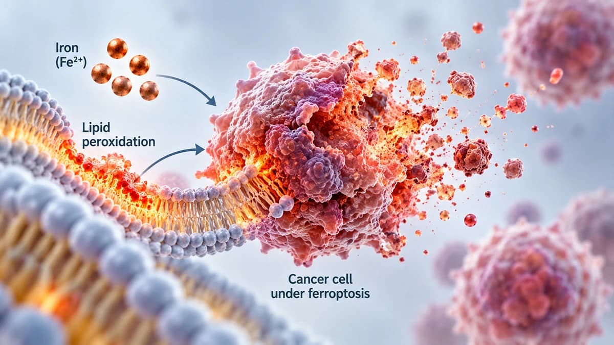
Every cell membrane contains fats, and some of those fats are more fragile than others. When a cell cannot keep this fragility in check, those fats start to break down through a chemical reaction called lipid peroxidation, essentially a slow-motion rusting of the cell's own membranes. Left unchecked, this damage spreads and eventually destroys the cell. This is what defines ferroptosis, and it looks nothing like other death pathways under a microscope. Apoptosis shrinks and neatly packages a dying cell for cleanup, using a cascade of cutting enzymes called caspases. Ferroptosis instead causes membranes to swell and eventually break apart, with none of the caspase activity apoptosis depends on. The distinction matters clinically, because many existing cancer treatments work by pushing cells toward apoptosis. Cells that have found ways to resist apoptosis might still be killed through this entirely separate route.
The Enzyme That Keeps Ferroptosis in Check
Cells are not defenseless against this kind of damage. A protective enzyme called GPX4 continuously repairs oxidized fats before they can accumulate, converting the toxic versions back into harmless ones. Think of GPX4 as a constant maintenance crew patching small leaks before they become a flood. When GPX4 is blocked or its levels drop, lipid peroxides build up quickly, and ferroptosis follows. This was shown directly in cancer cells, where knocking out or inhibiting GPX4 triggered ferroptotic death, making the enzyme one of the clearest levers researchers have found for switching this process on.
GPX4 cannot do its job alone. It depends on a small molecule called glutathione, the cell's primary antioxidant, to carry out its repair chemistry. Making glutathione requires a steady supply of an amino acid called cysteine, and cells import cysteine through a surface transporter known as system xc-, built around a protein called SLC7A11. Cancer cells that ramp up SLC7A11 activity keep their cysteine and glutathione supplies well stocked, which keeps GPX4 running and helps the cell resist ferroptosis. This transporter has become a focus of cancer research precisely because its activity seems to track with how resistant a tumor cell is to this death pathway.
Why Fat Composition Matters
Not all membrane fats are equally vulnerable. Polyunsaturated fatty acids, a category of fat with multiple weak chemical bonds, oxidize far more easily than more stable, saturated fats. An enzyme called ACSL4 determines how many of these fragile fats get built into a cell's membranes in the first place. Cells with high ACSL4 activity end up with membranes stocked full of oxidation-prone fat, which makes them considerably more sensitive to ferroptosis when their defenses falter. Cells with low ACSL4 activity, by contrast, tend to be comparatively protected, even under the same oxidative pressure. This helps explain why ferroptosis sensitivity varies so much from one cancer cell to another, independent of GPX4 levels alone.
Why Some Cancer Cells Seem Especially Exposed
One of the more striking findings in this field involves cells that survive cancer drug treatment. A small fraction of cancer cells can enter a temporary, slow-growing state after drug exposure, known as a drug-tolerant persister state, essentially hiding out rather than dying. Researchers working with Brent Stockwell's group found that these surviving cells, along with cancer cells in a more mesenchymal, migratory state, become unusually dependent on their lipid peroxidase defenses to stay alive. That dependency appears to make them more vulnerable to ferroptosis induction than the original, drug-sensitive tumor cells were. If confirmed more broadly, this would suggest a strategy for targeting exactly the cells that typically survive conventional treatment and go on to cause relapse, though this work currently rests on laboratory models rather than patient outcomes.
Ferroptosis also connects to one of cancer biology's most studied genes. TP53, a tumor suppressor that normally restrains cell growth and can trigger apoptosis, has also been shown to promote ferroptosis through a separate route that does not rely on its classical apoptotic signaling. This suggests TP53 may protect against cancer through more than one death pathway, widening the picture of how this well-known gene does its job.
A Possible Link to the Immune System
Another line of research connects ferroptosis to the immune system itself. During cancer immunotherapy, activated CD8+ T cells, the immune system's tumor-killing cells, release signaling molecules that can push tumor cells toward ferroptosis. This raises the possibility that part of immunotherapy's effect on tumors involves this iron-dependent death pathway, working alongside the immune system's more familiar killing mechanisms. The relationship is still being worked out, and it should be read as an active research direction rather than an established part of how current immunotherapies are understood to work.
Where the Research Stands
Taken together, these findings describe ferroptosis as a tightly regulated process sitting at the intersection of iron handling, fat chemistry, and the cell's antioxidant defenses, a connection that broader reviews of the field have mapped across metabolism and redox biology more generally. That breadth is part of why the topic has drawn sustained scientific interest: the same machinery shows up in contexts ranging from drug resistance to tumor suppression to immune signaling.
It is worth being precise about what this does and does not mean for patients today. No ferroptosis-inducing drug is currently an approved cancer treatment. The findings on persister cells, mesenchymal states, and immune-driven ferroptosis come primarily from cell culture and animal studies, not confirmed clinical results in people. None of this research supports using diet, iron restriction, or supplements as a way to trigger ferroptosis against cancer, and no current evidence backs that approach. What the research does offer is a genuinely new angle on an old problem: a death pathway separate from the ones most existing drugs rely on, and a growing map of what makes some cancer cells more exposed to it than others.
Common Questions
References
- Scott J. Dixon, Kathryn M. Lemberg, Michael R. Lamprecht, Rachid Skouta, Eleina M. Zaitsev, Caroline E. Gleason, Darpan N. Patel, Andras J. Bauer, Alexandra M. Cantley, Wan Seok Yang, Barclay Morrison, Brent R. Stockwell (2012). Ferroptosis: An Iron-Dependent Form of Nonapoptotic Cell Death
- Wan Seok Yang, Brent R. Stockwell (2016). Ferroptosis: Death by Lipid Peroxidation
- Wan Seok Yang, Rohitha SriRamaratnam, Matthew E. Welsch, Kenichi Shimada, Rachid Skouta, Vasanthi S. Viswanathan, Jaime H. Cheah, Paul A. Clemons, Alykhan F. Shamji, Clary B. Clish, Lewis M. Brown, Albert W. Girotti, Virginia W. Cornish, Stuart L. Schreiber, Brent R. Stockwell (2014). Regulation of Ferroptotic Cancer Cell Death by GPX4
- (2021). Cystine transporter SLC7A11/xCT in cancer: ferroptosis, nutrient dependency, and cancer therapy.
- Vasanthi S. Viswanathan, Matthew J. Ryan, Harshil D. Dhruv, Shubhroz Gill, Ossia M. Eichhoff, Brinton Seashore-Ludlow, Samuel D. Kaffenberger, John K. Eaton, Kenichi Shimada, Andrew J. Aguirre, Srinivas R. Viswanathan, Shrikanta Chattopadhyay, Pablo Tamayo, Wan Seok Yang, Matthew G. Rees, Sixun Chen, Zarko V. Boskovic, Sarah Javaid, Cherrie Huang, Xiaoyun Wu, Yuen-Yi Tseng, Elisabeth M. Roider, Dong Gao, James M. Cleary, Brian M. Wolpin, Jill P. Mesirov, Daniel A. Haber, Jeffrey A. Engelman, Jesse S. Boehm, Joanne D. Kotz, Cindy S. Hon, Yu Chen, William C. Hahn, Mitchell P. Levesque, John G. Doench, Michael E. Berens, Alykhan F. Shamji, Paul A. Clemons, Brent R. Stockwell, Stuart L. Schreiber (2017). Dependency of a therapy-resistant state of cancer cells on a lipid peroxidase pathway
- Matthew J. Hangauer, Vasanthi S. Viswanathan, Matthew J. Ryan, Dhruv Bole, John K. Eaton, Alexandre Matov, Jacqueline Galeas, Harshil D. Dhruv, Michael E. Berens, Stuart L. Schreiber, Frank McCormick, Michael T. McManus (2017). Drug-tolerant persister cancer cells are vulnerable to GPX4 inhibition
- Le Jiang, Ning Kon, Tongyuan Li, Shang-Jui Wang, Tao Su, Hanina Hibshoosh, Richard Baer, Wei Gu (2015). Ferroptosis as a p53-mediated activity during tumour suppression
- Weimin Wang, Michael Green, Jae Eun Choi, Miguel Gijón, Paul D. Kennedy, Jeffrey K. Johnson, Peng Liao, Xueting Lang, Ilona Kryczek, Amanda Sell, Houjun Xia, Jiajia Zhou, Gaopeng Li, Jing Li, Wei Li, Shuang Wei, Linda Vatan, Hongjuan Zhang, Wojciech Szeliga, Wei Gu, Rebecca Liu, Theodore S. Lawrence, Candice Lamb, Yuri Tanno, Marcin Cieslik, Everett Stone, George Georgiou, Timothy A. Chan, Arul Chinnaiyan, Weiping Zou (2019). CD8+ T cells regulate tumour ferroptosis during cancer immunotherapy
- (2017). ACSL4 dictates ferroptosis sensitivity by shaping cellular lipid composition.
- Brent R. Stockwell, José Pedro Friedmann Angeli, Hülya Bayir, Ashley I. Bush, Marcus Conrad, Scott J. Dixon, Simone Fulda, Sergio Gascón, Stavroula K. Hatzios, Valerian E. Kagan, Kay Noel, Xuejun Jiang, Andreas Linkermann, Maureen E. Murphy, Michael Overholtzer, Atsushi Oyagi, Gabriela C. Pagnussat, Jason Park, Qitao Ran, Craig S. Rosenfeld, Konstantin Salnikow, Daolin Tang, Frank M. Torti, Suzy V. Torti, Shinya Toyokuni, K.A. Woerpel, Donna D. Zhang (2017). Ferroptosis: A Regulated Cell Death Nexus Linking Metabolism, Redox Biology, and Disease
Team Review: This article has been reviewed by Canceria Research Team